LSP partners TESCAN to provide solutions in the field of SEM.
TESCAN is one of the global suppliers of scientific instruments. The company is building its reputation and brand name in the field of designing and manufacturing scanning electron microscopes and system solutions for different applications.
The company is focused on research, development and manufacturing of scientific instruments and laboratory equipment such as:
- scanning electron microscopes
- supplementary accessories for SEMs
- light optical microscopy accessories and image processing
- special vacuum chambers and custom systems
- detection systems
- scientific hardware and software development
TESCAN strives to keep improving continuously their products, which creates a competitive advantage for its customers.The TESCAN brand is becoming established thanks to the company’s participation in top research projects and cooperation with the leading companies in the field of electron microscopy and microanalysis. As a result, TESCAN’s instrumentation and innovative solutions have won a leading position in the world of nano- and microtechnology. Over 1200 SEM installations in more than 60 countries prove the highest technical solutions and first-class quality of TESCAN products.
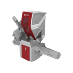
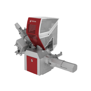
VEGA3 SB
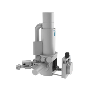
A fully PC controlled SEM with conventional tungsten heated cathode intended both for high vacuum as well as for low vacuum operations. Outstanding optical properties, flicker-free digital image with super clarity, sophisticated user-friendly software for microscope control and image capturing using Windows™ platform, standard formats of stored images, easy image management, processing and measurements, automatic setup of the microscope and many other automated operations are among the characteristic features of the equipment.
Analytical Potential
- The SB chamber is equipped with a 3-axis motorized stage
- First-class YAG scintillator-based detectors
- 10 chamber interface ports with optimized analytical geometry for e.g. EDX, EBSD, EBIC
- Selection of optional detectors and accessories
- Full operating vacuum can be reached within a few minutes with powerful turbomolecular and rotary fore vacuum pumps.
- Investigation of non-conductive samples in the variable pressure mode (SBU) version
- 3D measurements on a reconstructed surface utilizing 3D metrology software
VEGA3 SBU
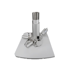
EasyProbe is a compact scanning electron microscope (SEM) fully integrated with a selected energy dispersive X-ray microanalyser (EDX). Superior imaging quality, high level of automation, easy usage and quick quantitative elemental results directly in the live image are among the characteristic features of the instrument. A variable pressure vacuum system allows investigation of non-conductive samples in their natural uncoated state.
Key Features of VEGA3 SB – EASYPROBE
- Fully integrated EDX microanalyser for automatic quantitative elemental analysis
- Original EasySEM software interface with One-Touch EDX toolbox
Microscope Control:
The EasySEM control interface is an easy-to-use mode of the main SEM control software. It is optimized for the use with a touchscreen. The EasySEM allows all basic imaging functions and a One-Touch EDX analysis toolbox, both with a great support of background automatics. When the One-Touch EDX tool is activated, the system automatically returns result of a quantitative elemental analysis of the area pointed in the live SEM image. The system allows advanced microscope control in three levels of user expertise. Control by the keyboard, the mouse and the trackball or optionally by the Control panel is available via the VegaTC control software using the Windows™ platform
VEGA3 LM
A fully PC controlled SEM with conventional tungsten heated cathode intended both for high vacuum as well as for low vacuum operations. Outstanding optical properties, flicker-free digital image with super clarity, sophisticated user-friendly software for microscope control and image capturing using Windows™ platform, standard formats of stored images, easy image management, processing and measurements, automatic setup of the microscope and many other automated operations are among the characteristic features of the equipment.
Analytical Potential:
- The LM chamber label indicates a large analytical chambere with a fully 5-axis motorized stage
- 11 chamber interface ports with optimized analytical geometry for EDX, WDX and EBSD
- First-class YAG scintillator based detectors
- Selection of optional detectors and accessories
- Full operating vacuum can be reached within a few minutes with powerful turbomolecular and rotary fore vackuum pumps
- Investigation of non-conductive samples in the variable pressure mode (LMU) version
- Several options of chamber suspension type ensure effective reduction of ambient vibrations in the laboratory
- 3D measurements on a reconstructed surface utilizing 3D metrology software
VEGA3 XM
A fully PC controlled SEM with conventional tungsten heated cathode intended both for high vacuum as well as for low vacuum operations. Outstanding optical properties, flicker-free digital image with super clarity, sophisticated user-friendly software for microscope control and image capturing using Windows™ platform, standard formats of stored images, easy image management, processing and measurements, automatic setup of the microscope and many other automated operations are among the characteristic features of the equipment.
Analytical Potential
- The XM chamber label indicates an extra large analytical chamber with a full 5-axis motorized stage with extended movements
- 12+ chamber interface ports with optimized analytical geometry for EDX, WDX and EBSD
- First-class YAG scintillator-based detectors
- Selection of optional detectors and accessories
- Full operating vacuum can be reached within a few minutes with powerful turbomolecular and rotary fore vacuum pumps.
- Investigation of non-conductive samples in the variable pressure mode (XMU) version
- Several chamber suspension type options ensure effective reduction of ambient vibrations in the laboratory.
- 3D measurements on a reconstructed surface utilizing 3D metrology software
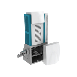
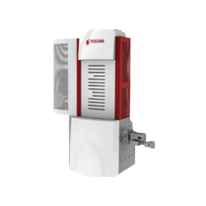
MIRA3 LM
A fully PC controlled FE SEM intended for both – for high vacuum as well as for low vacuum operations. Outstanding optical properties, flicker-free digital image with super clarity. Sophisticated user-friendly software for microscope control and image capturing using Windows™ platform, standard formats of stored images, enhanced image archiving, processing and measurements, automatic set up of the microscope and many other automated operations are characteristic features of the equipment.
Analytical Potential
- LM MIRA chamber provides superior specimen handling using a full 5-axis motorized compucentric stage and ideal geometry for EDX and EBSD.
- Numerous interface ports with optimized analytical geometry for EDX, WDX and EBSD as well as for attaching many other detectors
- First-class YAG scintillator-based detectors
- Selection of optional detectors and accessories
- Full operating vacuum can be obtained quickly and easily with powerful turbomolecular and dry fore vacuum pumps, electron gun pumping with an ion pump
- Investigation of non-conductive samples in variable pressure mode versions, excellent results in the investigation of magnetic samples
- Several options for chamber suspension type ensure effective reduction of ambient vibrations in the laboratory.
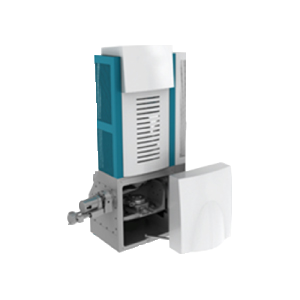
MIRA3 XM
The high performance MIRA3 XM. A fully PC controlled FE-SEM – for high vacuum as well as for low vacuum operations. Outstanding optical properties, flicker-free digital image with super clarity. Sophisticated user-friendly software for microscope control and image capturing using Windows™ platform. Standard formats of stored images, easy image management, processing and measurements, automatic set up of the microscope and many other automated operations are characteristic features of the equipment.
Analytical Potential
- XM MIRA chamber provide superior specimen handling using a full 5-axis motorized compucentric stage and ideal geometry for EDX and EBSD
- Numerous interface ports with optimized analytical geometry for EDX, WDX and EBSD as well as for attaching many other detectors
- First-class YAG scintillator-based detectors
- Selection of optional detectors and accessories
- Full operating vacuum can be obtained quickly and easily with powerful turbomolecular and dry fore vacuum pumps, electron gun pumping with an ion pump
- Investigation of non-conductive samples in variable pressure mode versions, excellent results in the investigation of magnetic samples
Several options for chamber suspension type ensure effective reduction of ambient vibrations in the laboratory
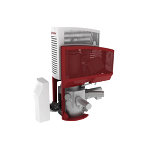
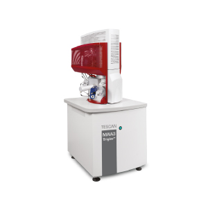
MAIA3 XM
A fully PC controlled ultra-high resolution FE-SEM intended for both – for high vacuum as well as for low vacuum operations. Outstanding electron-optical properties, flicker-free digital image with super clarity.Sophisticated user-friendly software for microscope control and image capturing using Windows ™ platform. Standard formats of stored images, easy image management, processing and measurements, automatic set up of the microscope and many other automated operations are characteristic features of the equipment.
Analytical Potential
- Extraordinary SE resolution with single pole 60⁰ objective lens
- Field-free mode for observing magnetic samples
- In-Beam SE Detector for inside the column detection of electrons at short working distances
- In-Beam BSE Detector for BSE imaging at very short working distances
- Full operating vacuum can be obtained quickly and easily with powerful turbomolecular and dry fore vacuum pumps, electron gun pumping with an ion pump
- Ideal geometry for EDX and EBSD
- Unique live stereoscopic imaging utilizing the 3D Beam Technology
User-Friendly Software and Software Tools
- Multi-user environment is localized in many languages.
- Four levels of user expertise/rights, including an EasySEM™ mode for quick routine investigations
- Image management and report creation
- Built-in self-diagnostics for system readiness checks
- Network operations and remote access/diagnostics
- Modular software architecture enables several extensions to be attached
LYRA3 XM
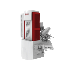
A fully PC controlled SEM with Schottky field emission cathode in combination with gallium Focused Ion Beam (FIB) column and optionally with Gas Injection System (GIS). Outstanding optical properties, flicker-free digital image with super clarity, sophisticated user-friendly software for SEM/FIB/GIS control and image capturing using Windows™ platform, standard formats of stored images, easy image management, processing and measurements, automatic set up of the system and many other automated operations are characteristic features of the equipment.
Analytical Potential
- XM chamber model provides superior specimen handling using a full 5-axis motorized compucentric stage and ideal geometry for EDX and EBSD
- Option of extra-large XM chamber with robust stage able to accommodate large samples
- Numerous interface ports with optimized analytical geometry for EDX, WDX and EBSD as well as for attaching many other detectors
- First-class YAG scintillator-based detectors
- Selection of optional detectors and accessories
- Full operating vacuum can be obtained quickly and easily
- Investigation of non-conductive samples in variable pressure mode versions, favorable conditions for the investigation of magnetic samples, non-distorted EBSD pattern compared to immersion magnetic lenses
Integrated active vibration isolation ensures effective reduction of ambient vibrations in the laboratory.
LYRA3 GM
A fully PC controlled SEM with Schottky field emission cathode in combination with gallium Focused Ion Beam (FIB) column and optionally with Gas Injection System (GIS). Oustanding optical properties, flicker-free digital image with super clarity, sophisticated user-friendly software for SEM/FIB/GIS control and image capturing using Windows platform, standard formats of stored images, easy image management, processing and measurements, automatic set up of the system and many other automated operations are characteristic features of the equipment.
Analytical Potential
- High brightness Schottky emitter for high-resolution / high-current / low-noise imaging
- Extraordinary resolution with powerful optional In-Beam SE Detector
- Unique three-lens Wide Field Optics™ design offering the variety of working and displaying modes embodying the TESCAN proprietary Intermediate Lens (IML) for the beam aperture optimization
- Real time In-Flight Beam Tracing™ for the performance and beam optimization integrating the well-established software Electron Optical Design. It includes also direct and continual control of beam spot size and beam current
- Fast imaging rate
- Beam Deceleration Technology (BDT) for excellent resolution at low beam voltages (optional)
- In-Beam BSE Detector for BSE imaging at very short working distances (optional) suitable even for ferromagnetic samples imaging
- High-throughput large-area automation, e.g. automated particle location and analysis
- Superior specimen handling using a motorized compucentric stage
- Ideal geometry for EDX and EBSD; non-distorted EBSD pattern
- Fast and easy obtaining of the clean chamber vacuum by powerful turbomolecular and dry fore vacuum pump; electron gun pumping by ion getter pump
- Fully automated microscope setup including electron optics setup and alignment
- Network operations and built-in remote access/diagnostics, all come as the TESCAN standard
- Unique live stereoscopic imaging utilizing the 3D Beam Technology
- Extended low vacuum mode with chamber pressure up to 500 Pa for non-conducting specimens imaging
- Unique ion optic column differentially pumped (2 ion getter pumps) for ultra-low ion scattering effect
- Motorized aperture changer in ion column with ultra-high reproducibility
- Beam Blanker and Faraday cup included as standard accessories for ion column
- Ultra-high resolution and excellent performance at high current with optional Cobra column
- Automatic FIB cutting and signal acquiring followed by 3D reconstruction (tomography), allowing 3D EBSD, 3D EBIC etc. with integrated 3D visualization
Sophisticated software for SEM/FIB/GIS control, image acquisition, archiving, processing and analysis; multi-user environment localized in many languages
GAIA3
TESCAN GAIA3 brings together an ultra-high resolution electron column and high-performance ion column fitted onto a single chamber. Built on the proven (MAIA3) FE-SEM platform, GAIA3 extends all of its qualities with the ability to modify surfaces with a focused ion beam. The outstanding imaging capabilities of the GAIA3 begin with its novel FE-SEM with excellent resolution at low voltages. The objective lens is narrower than a conventional objective and InBeam SE and InBeam BSE detectors placed in the column gives free space over the sample for unlimited FIB work and various analytical techniques.
Key Benefits
- Extraordinary SEM resolution with single-pole 60⁰ objective lens
- Best-in-class range of display modes- FIELD, RESOLUTION and DEPTH – based on TESCAN’s unique WIDE Field Optics
- Superior specimen handling using a motorized compucentric stage
- Field-free mode for observing magnetic samples
- High probe current, up to 200nA (SEM)
- E-beam lithography with ultra-short dwell time down to 20ns
- Ultra-high FIB resolution and excellent performance at high current with a Cobra column and probe current 1pA to 50nA
- Powerful DrawBeam toolbox including a number of basic and advanced programmable objects with various process parameters
- Besides the ability to investigate and modify the sample surface with enormous analytical potential, an extraordinary number of ports that enables all the detectors and technique to focus on a common analytical workspace.
General Benefits
The imaging, analysis and control of matter at the nanometer scale are key factors in successfully conducting research today. Resolution, accuracy, reproducibility, robustness and flexibility are key characteristics for a leading-edge tool.
Following three generations of success in electron microscopy, GAIA3 is the next generation platform introduced by TESCAN. Combining unsurpassed nanometer resolution SEM performance with excellent COBRA-FIB capabilities, the new TESCAN GAIA3 offers wide range of analytical compatibilities.
The greatest benefit of the GAIA3 is its low-voltage SEM imaging while maintaining a high resolution. This is essential especially for examining sensitive or non-conductive samples with a small interaction volume therefore giving excellent resolution and unique low voltage contrast.
COBRA-FIB is top-level technology in terms of resolution for both imaging and milling. It is the sharpest FIB instrument for nano-engineering in its class.
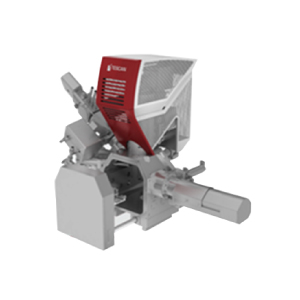
TESCAN Integrated Mineral Analyzer
TIMA, a fully automated, high throughput, analytical scanning electron microscope is designed specifically for the mining and minerals processing industry. The TIMA solution will address applications such as Mineral Liberation Analysis,TESCAN, a world leading manufacturer of scanning electron microscopes and focused ion beam workstations has introduced the TESCAN Integrated Mineral Analyzer.
TIMA, a fully automated, high throughput, analytical scanning electron microscope is designed specifically for the mining and minerals processing industry. The TIMA solution will address applications such as Mineral Liberation Analysis, process optimization, remediation, and search for precious metals and rare earths. TIMA measures modal abundance, size-by-size liberation, mineral association, and performs PGM search automatically on multiple samples of grain mounts and thin or polished sections.
TESCAN’s unique technology is based on a completely integrated EDX system that performs full spectrum imaging at very fast scan speeds. Image analysis in TIMA is performed simultaneously with SEM backscatter electron images and a suite of x-ray images. The level of hardware integration of the SEM and EDX allows for unprecedented acquisition speeds for fully automated data collection, resulting in fast, accurate, repeatable and reliable results. process optimization, remediation, and search for precious metals and rare earths
TIMA Advantages:
- Very fast and fully automated data acquisition process reached via SEM and EDX high level hardware integration
- System based on MIRA or VEGA SEM platform proven by customers in many countries
- Special VEGA column design significantly extending tungsten filament lifetime
- Newly designed exchangeable sample holder with integrated fixed BSE/EDX calibration standard and Faraday cup
- Possibility to modify size of samples according to customer demands
- Up to 4 integrated EDX detectors for maximum system performance
- New Peltier cooled EDX detector type for thermal stability guarantee
- Improved approach to data analysis increasing speed and reliability of process
- Variable dwell time and EDX analysis duration adapting to each part of the sample
- Software released in three editions
- Various modules for data analysis
- Customizable classification rules
- Favorable price to performance ratio
- Custom solution possibilities
FERA3 XM
A fully PC controlled SEM with Schottky field emission cathode in combination with Xe Plasma Focused Ion Beam (i-FIB) column and optionally with Gas Injection System (GIS). Outstanding optical properties, flicker-free digital image with super clarity, sophisticated user-friendly software for SEM/FIB/GIS control and image capturing using Windows™ platform, standard formats of stored images, easy image management, processing and measurements, automatic set up of the system and many other automated operations are characteristic features of the equipment.
Analytical Potential
- High brightness Schottky emitter for high-resolution / high-current / low-noise imaging
- Extraordinary resolution with powerful optional In-Beam SE Detector
- Unique three-lens Wide Field Optics™ design offering the variety of working and displaying modes embodying the TESCAN proprietary Intermediate Lens (IML) for the beam aperture optimization
- Real time In-Flight Beam Tracing™ for the performance and beam optimization integrating the well-established software Electron Optical Design. It includes also direct and continual control of beam spot size and beam current
- Fast imaging rate
- Beam Deceleration Technology (BDT) for excellent resolution at low beam voltages (optional)
- In-Beam BSE Detector for BSE imaging at very short working distances (optional) suitable even for ferromagnetic samples imaging
- High-throughput large-area automation, e.g. automated particle location and analysis
- Superior specimen handling using a motorized compucentric stage
- Ideal geometry for EDX and EBSD; non-distorted EBSD pattern
- Fast and easy obtaining of the clean chamber vacuum by powerful turbomolecular and dry fore vacuum pump; electron gun pumping by ion getter pump
- Fully automated microscope setup including electron optics setup and alignment
- Network operations and built-in remote access/diagnostics, all come as the TESCAN standard
- Unique live stereoscopic imaging utilizing the 3D Beam Technology
- Extended low vacuum mode with chamber pressure up to 500 Pa for non-conducting specimens imaging
- Unique ion optic column differentially pumped (2 ion getter pumps) for ultra-low ion scattering effect
- Motorized aperture changer in ion column with ultra-high reproducibility
- Beam Blanker and Faraday cup included as standard accessories for ion column
- Ultra-high milling rate and excellent performance at high currents
- Automatic FIB cutting and signal acquiring followed by 3D reconstruction (tomography), allowing 3D EBSD, 3D EBIC etc. with integrated 3D visualization
- Sophisticated software for SEM/FIB/GIS control, image acquisition, archiving, processing and analysis; multi-user environment localized in many languages
FERA3 GM
A fully PC controlled SEM with Schottky field emission cathode in combination with gallium Focused Ion Beam (FIB) column and optionally with Gas Injection System (GIS). Oustanding optical properties, flicker-free digital image with super clarity, sophisticated user-friendly software for SEM/FIB/GIS control and image capturing using Windows platform, standard formats of stored images, easy image management, processing and measurements, automatic set up of the system and many other automated operations are characteristic features of the equipment.
Analytical Potential
- High brightness Schottky emitter for high-resolution / high-current / low-noise imaging
- Extraordinary resolution with powerful optional In-Beam SE Detector
- Unique three-lens Wide Field Optics™ design offering the variety of working and displaying modes embodying the TESCAN proprietary Intermediate Lens (IML) for the beam aperture optimization
- Real time In-Flight Beam Tracing™ for the performance and beam optimization integrating the well-established software Electron Optical Design. It includes also direct and continual control of beam spot size and beam current
- Fast imaging rate
- Beam Deceleration Technology (BDT) for excellent resolution at low beam voltages (optional)
- In-Beam BSE Detector for BSE imaging at very short working distances (optional) suitable even for ferromagnetic samples imaging
- High-throughput large-area automation, e.g. automated particle location and analysis
- Superior specimen handling using a motorized compucentric stage
- Ideal geometry for EDX and EBSD; non-distorted EBSD pattern
- Fast and easy obtaining of the clean chamber vacuum by powerful turbomolecular and dry fore vacuum pump; electron gun pumping by ion getter pump
- Fully automated microscope setup including electron optics setup and alignment
- Network operations and built-in remote access/diagnostics, all come as the TESCAN standard
- Unique live stereoscopic imaging utilizing the 3D Beam Technology
- Extended low vacuum mode with chamber pressure up to 500 Pa for non-conducting specimens imaging
- Unique ion optic column differentially pumped (2 ion getter pumps) for ultra-low ion scattering effect
- Motorized aperture changer in ion column with ultra-high reproducibility
- Beam Blanker and Faraday cup included as standard accessories for ion column
- Ultra-high resolution and excellent performance at high current with optional Cobra column
- Automatic FIB cutting and signal acquiring followed by 3D reconstruction (tomography), allowing 3D EBSD, 3D EBIC etc. with integrated 3D visualization
- Sophisticated software for SEM/FIB/GIS control, image acquisition, archiving, processing and analysis; multi-user environment localized in many languages
XEIA3
New UHR SEM/FIB workstation
Whether your applications demand extremely powerful and ultra-fast micro-/nano-FIB machining, an outstanding image resolution at low beam energies, ultra- fast and reliable microanalysis or 3D analytical reconstructions, XEIA3 stands out as the ideal turnkey solution that offers all these capabilities in one single and unique instrument with ultimate performance.
KEY FEATURES
- Powerful SEM column equipped with a high brightness Schottky emitter for high currents, low-noise and extraordinary imaging
- In-Beam detectors for high signaling and excellent imaging at very shot working distances
- Ultra-fast xenon plasma ion source FIB. High Beam currents for outstanding milling speeds and an excellent performance in removing large volumes of material, and low beam currents for smooth polishing
- Less implantation, doping or degradation of insulator deposition a valuable feature for semiconductor industry
- Simultaneous SEM imaging during FIB milling or deposition (Two independent scan generators)
- Unique and advanced TESCAN’s technologies in terms of automated operations such as the In-flight Beam Tracing™ designed to accurately compute and adjust all the optimal parameters (WD, magnification, etc) for high resolution imaging
- Advanced patterning and 3D characterisations capabilities powered by DrawBeam, a pattern editing tool that also provided a real-time visualization during milling or lithographic processes
- Novel solution for fast 3-dimensional microanalysis such as 3D EDX and EBSD reconstructions
- Unique integration with TOF-SIMS and scanning probe microscope 12” wafer inspection by means of an extended chamber size for enabling 6”, 8” and 12” wafer inspection. 12” wafer inspection is an exclusive feature of TESCAN equipment
- Gas Injection System (GIS) for enhancing your FIB applications
- High-performance electronics for faster image acquisition up to 20ns/pxl, excellent deposition rate and an ultra-fast scannings
- Powerful turbomolecular and dry fore vacuum pump for keeping the chamber clean. Electron gun pumping by ion getter pump
Q-Phase
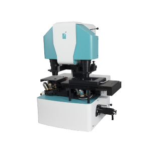
Tescan proudly introduces the Q-PHASE, a multimodal holographic microscope (MHM). With this instrument TESCAN expands in o the field of advanced light microscopy. The Q-Phase is a unique instrument for quantitative phase imaging (QPI) bases on the patented technology of Coherence-controlled holographic microscopy. This technology used incoherent light sources (Halogen lamp, LED) providing the highest quality, without any compromises and it is the only QPI technique enabling imaging of samples in scattering media. The Q-PHASE is purposely designed to observe living cells in vitro. It is based on a robust inverted transmission microscope platform. The whole system is situated in a microscope incubator. The full motorization fulfills even the highest demands regarding experiment automation. Futhermore, this system includes multiple imaging modes with fully integrated Fluorescence Module, simulated DIC and Brightfield imaging options. All these features make the Q-PHASE a valuable research tool for biological and biotechnical applications such as testing reactions of cells to a specific treatment-even with scattering non-transparent substances, monitoring cell’s life cycle including mitosis, distinguishing between different forms of cell deaths, analyzing cell growth, motility or morphology changes, imaging cells in extracellular matrices.
Key Features and advantages
- No image artifacts such as halo effect (as opposed to techniques based on Zernike phase contrast illumination)
- Enables very precise detection of cell boundaries
- Strong suppression of coherent noise(Speckles) & parasitic interferences (as opposed to laser-based approaches)
- Label-free- no staining is needed, simple sample preparation, observation of live cells in their native environment, no photobleaching problems
- Low phototoxicity- low light power density (10⁷x lower than fluorescence microscopy) allows long-term observations (for days)
- Coherence-gating effect- Q-PHASE special feature enabling to observe samples even in scattering media (phospholipid emulsions, extracellular matrices, etc)
- Multimodality- fully integrated fluorescence module, simulated DIC and brightfield which enables automatic multimodal imaging of the sample
- High- quality QPI- unique Q-PHASE’s optical setup allows using incoherent illumination which provides extraordinary imaging quality without any compromises
- Lateral resolution of conventional microscope (up to 2x better when compared to common laser-based approaches or pinhole spatial filtering based techniques)
- Fast acquisition- use of off-axis holographic approach makes Q-PHASE a single shot instrument, thus enabling imaging of very fast cell dynamics.
- Full motorization- focusing, sample stage, objective exchange, fluorescence filters
- Automated multidimensional acquisition- time-lapse, channel, position, Z-stack
- Simple image segmentation and processing – comparable to fluorescence date processing
- Quantitative- phase values can be recalculated e.g to cell dry-mass density (pg/um²) or direct topography with nanometer sensitivity (Usually non-biological samples with homogeneous refractive index distribution)
- High phase detection sensitivity- enables to detect even the smallest changes in axial direction, very sensitive detection of morphology or position changes